Key features and details | |
Cat. No. | MABL-3494 |
Name | Anti-CD40L [MR1](MABL-3494) |
Clone No. | MR1 |
From | Recombinant Antibody |
Isotype | Engineer antibody |
Application | Blocking, functional assays, FC |
Species Reactivity | Mouse |
Basic Information | |
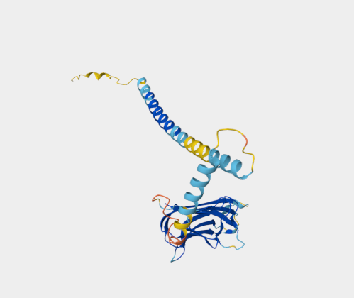
Specificity | This antibody is specific for murine CD40 ligand (CD40L), a 39 kDa transmembrane glycoprotein. CD40L is expressed transiently by activated T cells. Through its binding to CD40 on antigen presenting cells (APC) including B cells, monocytes/macrophages, and dendritic cells, it serves a crucial function in T cell-APC cognate interaction. CD40L-interaction with CD40 transduces signals for T-dependent B cell activation and induces B cells to enter the cell cycle. |
Alternative Name | CD154; CD40 ligand; CD40-L; Cd40l; gp39; HIGM1; IGM; IMD3; Ly-62; Ly62; RP23-153G22.3; T-BAM; T-cell antigen Gp39; TNF-related activation protein; Tnfsf5; TRAP; Tumor necrosis factor ligand superfamily member 5. |
UniProt | P27548 |
Immunogen | This antibody was raised by immunising Armenian hamsters with murine activated Th1 (D1.6) plasma membrane as described by Noelle et al (1992). |
Application Notes | This antibody was administred to SOD1 G93A mice, which exhibit ALS like symptoms, it was able to delay paralysis onset, it improved body-weight maintenance and extended survival (Lincecum et al., 2010; PMID:20348957). This antibody has been used in various FACS analyses for diverse immunological applications, such as to indicate how naive CD4 T cells constitutively express CD40L and augment autoreactive B cell survival (Lesley et al, 2006), to prove that the interaction between natural killer cells and dendritic cells has a pivotal role in the sensitization phase of contact hypersensitivity (Shimizuhira et al, 2014), and to demonstrate that enhanced CD8 T cell responses through GITR-mediated costimulation could resolve chronic viral infection (Pascutti et al, 2015). This antibody has also been used for in vitro and in vivo blocking and functional studies, for instance, to indicate that the 39-kDa CD40L membrane protein expressed on activated Th is a binding protein for CD40 and functions to transduce the signal for Th- dependent B-cell activation (Noelle et al, 1992), and to demonstrate that the CD40L–CD40 pathway can augment the survival of autoantigen-engaged B cells in the absence of T cell activation (Lesley et al, 2006). |
Antibody First Published | Noelle et al. A 39-kDa protein on activated helper T cells binds CD40 and transduces the signal for cognate activation of B cells. Proc Natl Acad Sci U S A. 1992 Jul 15;89(14):6550-4. PMID:1378631 |
Note on publication | Describe the original generation of this antibody and its subsequent use in FACS, blocking and functional studies to indicate that the 39-kDa membrane protein expressed on activated Th is a binding protein for CD40 and functions to transduce the signal for Th-dependent B-cell activation. |
COA Information (For reference only, actual COA shall prevail) | |
Size | 100 μg Purified antibody. |
Concentration | 1 mg/ml. |
Purification | Protein A affinity purified |
Buffer | PBS with 0.02% Proclin 300. |
Concentration | 1 mg/ml. |
Storage Recommendation | Store at 4⁰C for up to 3 months. For longer storage, aliquot and store at - 20⁰C. |


