Product Name
Mouse IgG1 Fab and F(ab')2 Preparation Kit
Size
10 samples for 250ug-4mg IgG
Principle
Immobilized Ficin
Description
Mabioway Mouse IgG1 Fab and F(ab
Features
• High capacity—kit provides for fragmenting 0.25 to 4 mg IgG.• Complete—kit includes all reagents needed to prepare and purify antibody fragments• Enzyme-free digestion products—Immobilized Ficin (beaded agarose resin) provides for control of the digestion reaction and complete removal of resulting antibody fragments from the proteolytic enzyme• Optimized for mouse IgG1—the kit procedure is optimized for mouse IgG1; the only effective method for this IgG species and subclass
Application
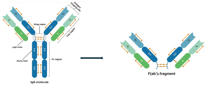
Mouse IgG1 Fab and F(ab´)2 Preparation Kit uses Immobilized Ficin to prepare fragments from mouse IgG1. Ficin generates F(ab´)2 fragments exclusively in the presence of 1-4mM cysteine; Fab fragments are generated in the presence of 25mM cysteine (Figure 1). Fragment generation from other IgG species and isotypes might be possible by modifying the cysteine concentration and other digestion parameters.Pepsin is commonly used for generating F(ab´)2 fragments because the pepsin cleavage site on human IgG contains a Leu 234, which is conserved in most species; however, mouse IgG1 lacks this residue and others, which possibly contributes to the restricted hinge region and resistance to pepsin cleaveage. Also, the low pH required for pepsin digestion can destroy or damage antibodies. For comparison, mouse IgG1 monoclonal antibodies were digested with ficin, pepsin, bromelain and elastase. Ficin digestion produced high yields of F(ab´)2 fragments with the highest residual antigen-binding activity and immunoreactivity. Affinity constants of ficin-generated F(ab´)2 fragments were near those of intact antibody.This kit contains the necessary components for Fab or F(ab´)2 generation of mouse IgG1 and subsequent purification. Immobilized Ficin enables immediate cessation of the digestion by simply removing the resin from the antibody digest solution. The included Columns allow easy manipulation of the resin and maximum Fab and F(ab´)2 recoveries. The prepacked, immobilized Protein A Column and optimized binding buffer binds the intact Fc fragments and undigested IgG, allowing for efficient Fab or F(ab´)2 fragments purification. The optimized cysteine concentration produces Fab or F(ab´)2 with maximum purity. This complete kit makes Fab and F(ab´)2 generation and purification simple, fast and effective.
Product Information
• These instructions are optimized for mouse IgG1. Fragmentation of other mouse IgG isotypes or IgG from other species might require optimization.• The kit components and protocol are configured for 0.5mL samples containing 0.25-4mg of IgG. For 25-250µg samples, use the Mouse IgG1 Fab and F(ab´)2 Micro Preparation Kit (Product No. MAK-0005).• Proper sample preparation is essential for successful fragment generation using this kit. If the IgG sample contains a carrier protein such as BSA, use the Antibody Clean-up Kit (Product No. MAK-0007) to remove it before performing the buffer exchange (Section B).• Protein A Binding Buffer will precipitate in SDS-PAGE Loading Buffer. Dilute sample 1:5 or desalt or dialyze before loading onto a gel.
Contents
10 antibody samples, each containing 0.25 to 4 mg IgG• Immobilized Ficin Agarose, 1.5 mL• Cysteine-HCl, 0.5g• Mouse IgG1 Digestion Buffer, 100 mL• Protein A Column, 1 mL, 1 column• Protein A Binding Buffer, 100 mL• IgG Elution Buffer, 100 mL• Desalt Columns, 2 mL, 10 columns• Microcentrifuge tubes, 2 mL, 30 tubes
Additional Materials
• Incubator capable of maintaining 37°C• Microcentrifuge capable of 5,000 × g• Variable speed centrifuge• 15mL conical collection tubes• End-over-end mixer or tabletop rocker• 0.02% Sodium azide storage solution (in PBS or TBS) for the Immobilized Protein A
Material Preparation
Digestion BufferFab generation: Dissolve 43.9mg cysteine•HCl in 10mL of the supplied Mouse IgG1 Digestion Buffer. After adding the cysteine•HCl the pH should be ~5.6.F(ab´)2 generation: Dissolve 7mg cysteine•HCl in 10mL of the supplied Mouse IgG1 Digestion Buffer. After adding the cysteine•HCl the pH should be ~5.9.Note: Cysteine readily oxidizes to cystine; therefore, prepare this buffer on the same day of use.
Procedure
A. Immobilized Ficin Equilibration1. Gently swirl the Immobilized Ficin vial to obtain an even suspension. Seat the spin-column frit with an inverted 200µL pipette tip.2. Tplace place 100µL of the 50% slurry (i.e., 50µL of settled resin) into the 1.5mL column. Centrifuge the column at 5000 × g for 1 minute and discard buffer.3. Wash resin with 0.15mL of Digestion Buffer. Centrifuge column at 5000 × g for 1 minute and discard buffer. B. IgG Sample Preparation1. Desalting Column and loosen cap. Place column in a 15mL collection tube.2. Centrifuge column at 1500 × g for 1 minute to remove storage solution. Place a mark on the side of the column where the compacted resin is slanted upward. Place column in centrifuge with the mark facing outward in all subsequent centrifugation steps.Note: Resin will appear compacted after centrifugation.3. Add 500µL of Digestion Buffer to column. Centrifuge at 1500 × g for 1 minute to remove buffer. Repeat this step three additional times, discarding buffer from the collection tube.4. Place column in a new collection tube, apply 500µL of sample to the center of the compacted resin bed.5. centrifuge at 1500 × g for 2 minutes to collect the sample. Discard the column after use.6. If IgG sample is 0.5-8mg/mL (i.e., 250µg to 4mg), no further preparation is necessary. If sample volume is less than 500µL, add Digestion Buffer to a final volume of 500µL.C. Generation of Fragments1. Add 500µL of the prepared IgG sample to the column tube containing the equilibrated Immobilized Ficin. Place top cap and bottom plug on column.2. Incubate digestion reaction 3-5 hours for generation of Fab fragments or 24-30 hours for generation of F(ab´)2 fragments with end-over-end mixer or tabletop rocker at 37°C. Maintain constant mixing of resin during incubation.3. Remove the bottom cap and place the column into a 2mL microcentrifuge tube. Centrifuge the column at 5000 × g for 1 minute to separate digest from the Immobilized Ficin.4. Wash resin with 500µL PBS. Place column into a 2.0mL microcentrifuge tube. Centrifuge column at 5000 × g for 1 minute. Repeat this step for a total of three washes.5. Add the wash fractions to the digested antibody from Step 3. Total sample volume should be 2mL. Discard used Immobilized Ficin.Note: To assess digestion completion, evaluate the digest and wash fraction via SDS-PAGE. Dilute digest 1:5 before adding to SDS-PAGE loading buffer. Because of the presence of cysteine, boiling samples in non-reducing SDS-PAGE loading buffer will reduce the sample. To avoid reducing the 50kDa Fab fragment or 110kDa F(ab´)2 fragment do not boil the samples. For best interpretation, desalt or dialyze samples before electrophoresis. See representative gels in the Additional Information Section.D. Fab and F(ab´)2 Purification1. Equilibrate the Protein A Column, Protein A Binding Buffer and IgG Elution Buffer to room temperature. Set centrifuge to 1000 × g.2. Place column in a 2mL collection tube and centrifuge for 1 minute to remove storage solution (contains 0.02% sodium azide). Discard the flowthrough.3. Equilibrate column by adding 500µL of Protein A Binding Buffer and briefly mix. Centrifuge for 1 minute and discard the flow-through. Repeat this step once.4. Add the digested antibody sample (Step C.5) to column and tightly cap top. Suspend resin and sample by inversion. Incubate at room temperature with end-over-end mixing for 10 minutes.5. Place column in a new 15mL collection tube and centrifuge for 1 minute. Save the flow-through as this fraction contains Fab and F(ab´)2 fragments.6. For optimal recovery, wash column with 500µL of Protein A Binding Buffer. Centrifuge for 1 minute and collect the flow-through. Repeat and combine wash fractions with the Fab or F(ab´)2 fraction from Step 5.7. Apply 500µL of IgG Elution Buffer to the Protein A Column and centrifuge for 1 minute. Repeat this step two times to obtain three fractions, which will contain undigested IgG and Fc fragments. To save the undigested IgG or Fc fragments, add 40µL of a neutralization buffer (e.g., 1M phosphate or 1M Tris at pH 8-9) to each elution fraction.8. Estimate protein concentration by measuring the absorbance at 280nm. Use an estimated extinction coefficient of 1.4. Alternatively, measure the concentration using the Reducing Agent Compatible BCA Protein A ssay; however, the sample must contain less than 2.5mM cysteine. The combined digest and Protein A fraction might contain up to 2mM cysteine. The Protein A Binding Buffer might also interfere with colorimetric Protein A ssays. For best results, desalt or dialyze the sample and use the Reducing Agent Compatible BCA Protein A ssay.E. Regeneration of the Immobilized Protein A Column1. Add 1mL of IgG Elution Buffer and centrifuge for 1 minute. Discard flow-through and repeat once.2. Add 1mL of PBS to the column, centrifuge for 1 minute and discard the flow-through. Repeat three times.3. For storage, add 400µL of 0.02% sodium azide in PBS to the column. replace top and bottom caps. Store column upright at 4°C. Columns can be regenerated at least 10 times without significant loss of binding capacity.
Trouble shooting
Problem 1: Low amounts of Fab (50kDa) or F(ab´)2 (110kDa) produced as visualized by non-reducing SDS-PAGE1)IgG sample was not prepared properly.Buffer exchange IgG into Digestion Buffer.2)Sample loading buffer contains reducing reagent.Use SDS loading buffer that does not contain β-mercaptoethanol, DTT or TCEP.3)Digested material contains cysteine.Desalt digest before SDS-PAGE.4)Sample contains protein other than IgG (e.g., BSA).Remove BSA with the Antibody Clean-up Kit.5)Some mouse IgG1 clones may generate alternate fragments of different molecular weightUse the Fab Preparation Kit (Product No. 44985) or F(ab´)2 Preparation Kit (Product No. 44988) and dilute mouse IgG1 samples with Protein A Binding Buffer (Product No. 21001) for purification.Problem 2: Fab or F(ab´)2 has low immunoreactivitySample digested for too long. Reduce digestion time; do not exceed 8 hours for Fab or 40 hours for F(ab´)2 or try using the F(ab´)2 or Fab Preparation Kit.Problem 3: Protein A flow-through contains Fab and F(ab´)21)Extended digestion times for F(ab´)2 production might result in the formation of Fab.Use recommended cysteine concentrations and digestion times.
 MAK-0006-Mouse IgG1 Fab and F(ab')2 Preparation Kit.pdf
![]() MAK-0006-Mouse IgG1 Fab and F(ab')2 Preparation Kit.pdf
MAK-0006-Mouse IgG1 Fab and F(ab')2 Preparation Kit.pdf
![]() MAK-0006-Mouse IgG1 Fab and F(ab')2 Preparation Kit.pdf
MAK-0006-Mouse IgG1 Fab and F(ab')2 Preparation Kit.pdf
